

⚡ Article Synopsis: In this article, image analysis scientist Matthew McFarlane shares his thoughts on some of the recent Visiopharm software updates, how they work, and their benefits.
With the rapid advancement into the era of precision medicine, digital pathology is proving to be an invaluable development in answering biological questions that have remained a mystery up until now.
To accurately capture the molecular underpinnings of complex pathologies, the staining and scanning of histological samples have shown significant improvements in recent years, with some technologies now able to generate ultra-high-plex imaging data.
In order to keep up with the increasing complexity of multiplex immunofluorescence (mIF) images, OracleBio is continually evaluating new image analysis tools and implementing these into our workflows. One such software offering robust yet versatile tools to analyse traditionally challenging images is the Visiopharm platform.
With the most recent update to the platform (version 2023.01), we have been eager to highlight some of the new and optimized features that will help derive valuable and accurate data from your ultra-high-plex images.
Advanced Colour Adjustment Menu
A significant change in the latest production release of Visiopharm is the advanced colour adjustment menu. To explore the complex interactions occurring in biological systems, ultra-high-plex images can become increasingly challenging to handle and visualise. To improve upon this, Visiopharm has redesigned the advanced colour adjustment menu to enhance accessibility and management of image channel settings.
Additionally, with the increasing popularity of combining core marker panels in a single staining protocol – such as tumour, immune, or structural profiling panels – the colour adjustment menu now allows for channel grouping. With the ability now to create custom groups that can mirror the staining profiles applied to the tissue, image visualisation becomes considerably more adaptable to the needs of the image analyst. Lastly, these colour settings and group structures can now be saved to configurations, allowing for them to be quickly applied to all images in your study or across batches.
Phenoplex Guided Workflow
A second major update to the platform was the addition of the Phenoplex Guided Workflow. With this new intuitive module, cell phenotyping of high-plex images has become significantly more manageable with easily adjustable thresholds updated in real time. Once thresholds have been set for all biomarkers of interest, Visiopharm has implemented various visualisation tools to verify the phenotype results from your images.
Using the co-occurrence matrix, threshold settings can be assessed by reviewing the number of cells positive for all selected biomarker combinations. This can be helpful in identifying any unexpected biomarker co-expression counts, where the interactive plot can now highlight specific cell phenotype populations in the image to examine whether thresholds require adjustment.
In addition, the phenotype list tool enables quick and easy review of all cell phenotypes and selectively toggle their visibility. In combination with the colour adjustment menu updates, verifying increasing numbers of complex phenotypes has become a much more manageable and user-friendly task.

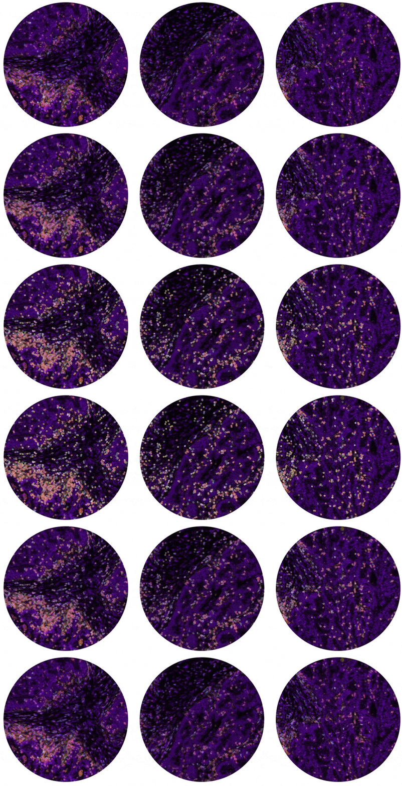
Figure 1: Visiopharm’s Phenoplex Guided Workflow. A) Using the phenoplex guided workflow, the user can select biomarkers of interest to threshold and Visiopharm will create overlays for all cellular phenotypes detected. Phenoplex overlays can then be toggled to view individual or a combination of cellular phenotypes. Here, a custom immune panel was selected as shown in the phenotype list on the left, and colour management group settings on the right. B) Higher magnification examples of the Phenoplex overlays for all phenotypes generated as shown in part A.
Overall, the regular updates to the Visiopharm platform help keep OracleBio at the forefront of quantitative digital image analysis.
These novel tools are critical in maintaining and improving the accuracy and productivity of our image analysis team in this exciting progression into ultra-high-plex image analysis.
With plenty more valuable updates to the platform on the horizon, our image analysis capabilities are continually adapting and evolving to meet your analytical needs.
If you’d like to learn more about how our scientists utilise Visiopharm or how we can support your image analysis studies, please don’t hesitate to get in touch with us here.
How OracleBio can support you
Why work with OracleBio?
Stay up to date with OracleBio
Sign up to our mailing list to stay informed about news, webinars, events and more