
⚡ Article Synopsis: In this article, OracleBio Senior Clinical Pathologist, Dr. James Going, discusses the history of microscopy and his experiences throughout his career; exploring how advancements in technology have both challenged and strengthened the field, and examining the ongoing debate about the role of microscopy and digital pathology in modern medicine.
Laying the Foundations
It is widely accepted that history goes in cycles, and this can be true for science too.
Sometimes, things from the past come back in a new form. When a new methodology appears, we are excited by its newness, then as the newness wears off, we begin to realise that the marvelous new ‘thing’ is not as marvelous as we had dreamed or hoped.
With time, we come to a more balanced appreciation of the strengths of the new technology and begin the long work of integrating old and new into a stronger unity.
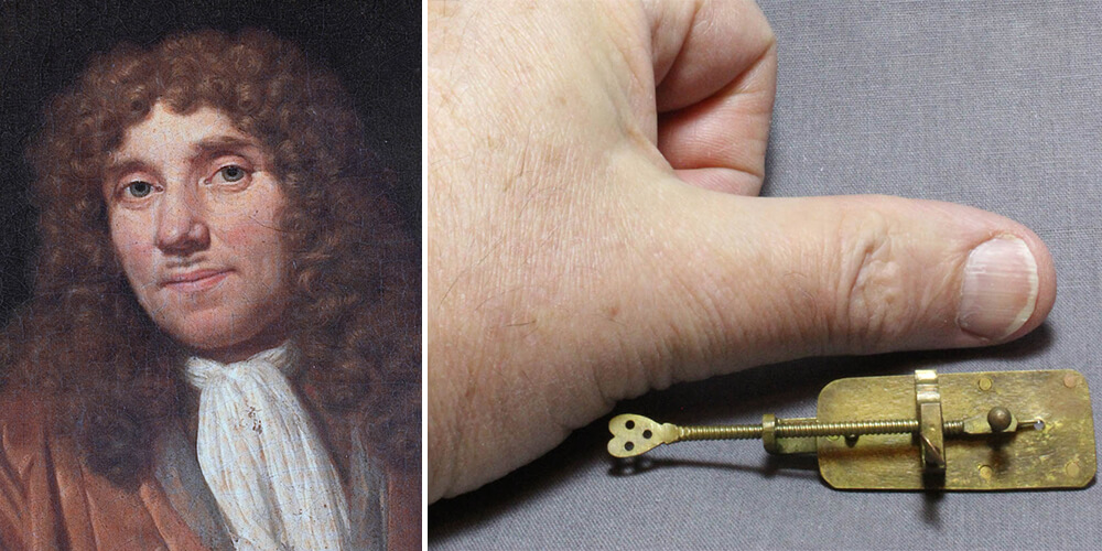
[Above]: Antonie Leeuwenhoek 1632-1723: the first person to see bacteria and protozoa, using microscopes he made himself – pictured.
Microscopy is around four centuries old but had to wait half a century for the first really significant microscopist – Antonie Leeuwenhoek (pictured above), in the second half of the seventeenth century.
But as early as the first quarter of the eighteenth century, people were beginning to say that microscopy was démodé, outdated, passé.

[Above]: Ernst Abbe 1840-1905: a physicist who perfected the optical microscope, in collaboration with Carl Zeiss and Otto Schott.
In the nineteenth century, there were important technical advances. The invention of the achromatic microscope by Joseph Jackson Lister was followed by more highly corrected apochromatic lenses developed by optician and physicist Ernst Abbe (pictured above).
These optical achievements and developments in microtomy revitalised microscopy, but after only a few years the cycle would be repeated. German microbiologist Paul Ehrlich, in the early twentieth century, could again suggest that microscopy was outmoded.
In the mid-twentieth century, American physician, Albert Coons developed immunofluorescence, which with the later work of César Milstein and Georges Köhler on monoclonal antibodies, initiated a further genuine revolution in histology.
Before long, though, around the turn of the 20th and 21st centuries, advancing technologies and discoveries involving DNA and RNA led some people to think (once again) that microscopy in general and diagnostic histopathology based on morphology were outdated; even that the latter might disappear altogether as a living scientific discipline.
A particular criticism has been that microscopy shows us things but does not tell us how or why. Mechanistic explanations have been held to be the business of other sciences, by implication more powerful and more serious than microscopy and histology.
In histopathology, the pejorative term ‘stamp collecting’ was sometimes encountered. And yet, if we are ignorant of the phenomena to be explained, the work of explanation cannot begin! Microscopy alone does not tell us everything we need to know, but without it, we are lost.
Digital Democratisation of Pathology
We can say for certain, then, that histology and histopathology resting on morphological foundations will be with us for the foreseeable future.
Equally certain is that how we practice diagnostic histopathology will change. Technologies that are going to be part of that change are already with us, and fairly mature. Digital pathology has been a long time coming, but it is here now.
Digital pathology liberates the histological image from the microscope and the laboratory so that the same image can be interpreted as easily by an observer, not necessarily a doctor, on the other side of the world.
This liberation makes possible what we may call the democratisation of pathology. Up to now diagnostic histopathology has usually been practiced by medical experts after lengthy training (10+ years). Medical understanding of the ‘fuzzy logic’ of clinical reasoning and the Bayesian character of diagnostic inference has been important, but, there is a worldwide shortage of doctors, including histopathologists.
Medical school education once involved relatively high exposure to histology and histopathology, allowing students with an affinity for the interpretation of histological imagery to discover that affinity. The decreased emphasis in recent years on histology and histopathology in medical education seems likely to reduce the supply of medical histopathologists for the future. We could see this as a threat, or take a more positive outlook and see it as an opportunity to do things differently.
‘Digital democratisation’ has the potential to make histology and histopathology more generally accessible to people with expertise other than those traditionally associated with diagnostic histopathology.
Looking Ahead: New Partnerships

[Above]: An example of the importance of computer science in supporting innovation can be highlighted by Kathleen Booth (1922-2022), a British computer scientist and co-designer of one of the world’s first operational computers. She invented and wrote the first assembly language, which led to powering the world’s first supercomputers used in scientific research. Booth was also a pioneer of neural networks in biology.
There are many precedents for tasks once regarded as the exclusive province of the doctor successfully being adopted by other professions. Nurses now perform endoscopies and some surgical procedures.
Some things for which pathologists have classically been responsible, such as breast cancer and prostate cancer grading, are attractive candidates for early integration with digital pathology. The necessity for the participation of computer scientists and image analysts as equal partners in the development and integration of digital pathology in diagnostic care is obvious. Indeed, I see similarities with the integration of clinical pathologist expertise in digital pathology R&D and drug development projects, where digital pathology is more established and driven by image analysis scientists.
I look forward to a broader spectrum of medical and non-medical pathologists working together to establish the diagnostic tools of the future, in which computational (digital) pathology will play a central part. There will be challenges to overcome, but no change is not an option in a changing world!
In my next blog, I will discuss how, as a clinical anatomical pathologist, I work alongside image analysis scientists and computer scientists at OracleBio in developing workflows to accurately detect cells and morphological features within tissue samples from R&D studies.
References
- Greenough RB. Varying degrees of malignancy in cancer of the breast. J Cancer Res 1925;9:425–463
- Patey DH, Scarff RW. The position of histology in the prognosis of carcinoma of the breast. Lancet 1928;1:801–804
- Elston CW, Ellis IO. Pathological prognostic factors in breast cancer. The value of histological grade in breast cancer: experience from a large study with long-term follow-up. Histopathology 1991;19:403–410

About the author:
Dr. James Going – Senior Clinical Pathologist at OracleBio
With over 39 years of pathology experience as a Clinical Pathologist in the NHS, James has a wide and deep knowledge of diagnostic histopathology with specialist expertise in breast and gastrointestinal tract.
Find out more about OracleBio's Pathology Expertise
OracleBio's Quantitative Digital Pathology Services
Stay up to date with OracleBio
Sign up to our mailing list to stay informed about news, webinars, events and more