
Angela Vasaturo, PhD; Senior Scientist, Ultivue
Lorcan Sherry, PhD; Chief Science Officer, OracleBio
Broadcast via The Pathologist, November 2020
Multiplex staining and image analysis are being increasingly deployed for immuno-oncology (I/O) evaluations to investigate and unravel the complexity of immune cell/tumor cell interactions in the tumor microenvironment (TME) and in discovering novel predictive biomarkers for targeted immunotherapy. Immune checkpoint inhibitor treatment, guided by PD-L1 and PD-1 tissue expression assessed by immunohistochemistry, has already shown to improve outcomes for non-small cell lung carcinoma (NSCLC) and colorectal cancer (CRC) metastatic patients. During this webinar, we’ll present a workflow utilizing an 8-plex staining protocol and detailed image analysis approach for the comprehensive assessment of the immune cell types and interactions within the TME, including co-localization and spatial phenotyping analysis in NSCLC and CRC tumor types.
Learning Objectives:
- A fast, optimized and validated 8-plex multiplex immunofluorescence staining protocol and a comprehensive image analysis can be applied to a broad range of cancer types in order to characterize the immune contexture.
- The ability to carry out multiparameter cellular analysis facilitates the delivery of key cell phenotypes as well as their functional status, thereby leading to greater understanding of tumor biology.
More from OracleBio:
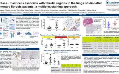
MedImmune Poster Presentation, European Respiratory Society 2016
OracleBio were delighted to present a poster at the Eropean Respiratory Society (ERS) Conference, London, Sept, 2016. The poster, entitled ‘Tryptase+ mast cells associate with fibrotic regions in the lungs of idiopathic pulmonary fibrosis patients; a multiplex...
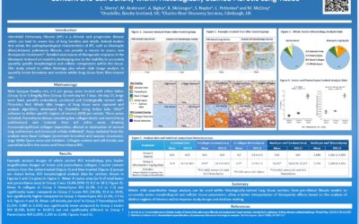
Whole Slide Lung Tissue Image Analysis Poster Presentation, European Respiratory Society 2016
OracleBio were delighted to present a poster at the Eropean Respiratory Society (ERS) Conference, London, Sept, 2016. The poster, entitled ‘Utilisation of Whole Slide Image Analysis to Quantify Lesion Formation, Collagen Content and Cell Density within Histologically...
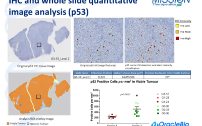
OracleBio Xenograft IHC biomarker image analysis data presented at the Tumour Models Conference, London December 2014
The use of histology image analysis to accurately quantify biomarker levels in oncology research is a key approach used to better understand the in vivo drug response and mechanism of action of new therapies. Whole slide image analysis can help to extract detailed,...
Find out more about our Multiplex IF Analysis
OracleBio's Quantitative Digital Pathology Services
Stay up to date with OracleBio
Sign up to our mailing list to stay informed about news, webinars, events and more