
CD8 Analysis in NSCLC Tumour Tissue Using HALO

Study Overview
This case study aimed to quantify CD8+ immune cells within the tumour microenvironment and invasive margin regions of IHC-stained NSCLC clinical tumour tissue, and to assess the spatial distribution of CD8+ cells across the tumour microenvironment.
The workflow used image analysis to generate total immune cell counts in tumour and stroma regions, immune cell ratios across tumour-stroma-invasive margin regions, immune-tumour cell proximity analysis, and immune cell infiltration readouts. HALO was used to develop deep learning classifiers for tissue classification and invasive margin analysis, as well as nuclear segmentation and cell analysis modules for CD8-positive and negative cell detection.
Key outputs included CD8+ cell counts, normalised ratio metrics, infiltration band analysis, and proximity-based spatial analysis. The study demonstrated an end-to-end collaborative workflow between IQVIA and OracleBio for transforming NSCLC clinical tumour tissue into quantitative pathology data.
Sample Details
NSCLC clinical tumour tissue.
Staining
IHC staining for CD8.
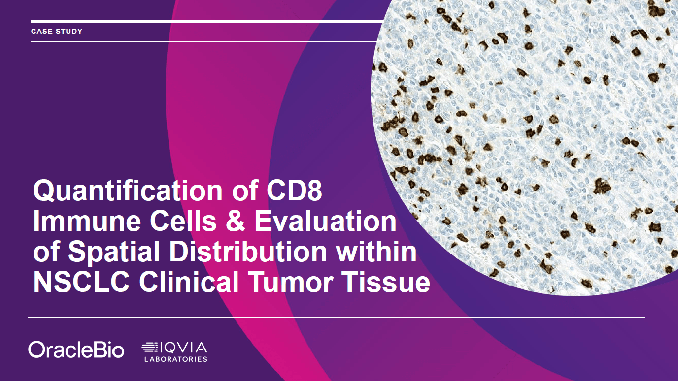
CD8 IHC-positive cells shown with brown staining.
Analysis
Image analysis performed using HALO.
Deep learning classifiers developed in HALO for tumour, stroma, tissue, glass, white space, necrosis, and other tissue classes.
CD8-positive and negative cell detection performed using Nuclear Segmentation AI and Cell Analysis Modules in HALO.
View the Case Study
Click the thumbnail below to view the case study PDF:
Benefits of Working with OracleBio for your Clinical Studies:
- Data you can trust – We have thorough quality control procedures in place throughout our workflow to ensure the data we provide is of a high-quality standard.
- Work alongside Project managers – Experienced with ensuring data parity between time-separated batches of clinical samples.
Note: Permission has been granted for the use of this material