
Download the PDF poster:

Watch the presentation on-demand:
Presented at Digital Pathology & AI Congress USA 2025 | In support of the INCISE Project
As part of the INCISE project, an ambitious collaboration between the University of Glasgow, NHS Greater Glasgow and Clyde, and a network of industry partners, OracleBio led the Digital Pathology work package, helping to advance bowel cancer surveillance through scalable image analysis.
Our role focused on developing and deploying an automated pipeline to quantify Ki67 biomarker expression across a large cohort of colorectal polyp samples. This data forms part of a wider effort to create a multi-omics risk model that can help stratify patients and personalise future screening pathways.
What you’ll learn:
- A four-stage AI-driven image analysis workflow – Built in Visiopharm and validated by expert pathologists, our pipeline included tissue QC, adenoma classification, gland segmentation, and single-cell phenotyping.
- Execution at scale – The final pipeline was used to process over 2,200 Ki67-stained whole slide images, executed in parallel across 75 GPU-enabled cloud nodes.
- Fast, reliable performance – All 2,211 samples were analysed in just 45 hours—demonstrating the pipeline’s robustness, scalability, and future potential to support high-throughput diagnostic workflows.
Click the thumbnail above to download the full poster and see how OracleBio’s image analysis expertise is helping power the next generation of bowel cancer surveillance.
Stay up to date with OracleBio
Sign up to our mailing list to stay informed about news, webinars, events and more
You might also like these Posters:
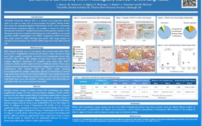
Whole Slide Lung Tissue Image Analysis Poster Presentation, European Respiratory Society 2016
OracleBio were delighted to present a poster at the Eropean Respiratory Society (ERS) Conference, London, Sept, 2016. The poster, entitled ‘Utilisation of Whole Slide Image Analysis to Quantify Lesion Formation, Collagen Content and Cell Density within Histologically...
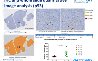
OracleBio Xenograft IHC biomarker image analysis data presented at the Tumour Models Conference, London December 2014
The use of histology image analysis to accurately quantify biomarker levels in oncology research is a key approach used to better understand the in vivo drug response and mechanism of action of new therapies. Whole slide image analysis can help to extract detailed,...