

⚡ Article Synopsis: OracleBio’s Matthew Corser explains how to build an accurate AI-powered tissue classifier for IHC-stained skin samples using HALO AI’s DenseNet v2 network, sharing the key practical insights needed to achieve precise, generalisable segmentation of skin tissue layers across a full preclinical study cohort.
The ability to quantify biomarker staining within specific regions of skin tissue provides a deeper understanding of spatial expression and enables a more accurate interpretation of therapeutic efficacy. In this post, we share the key insights gained from a recent preclinical imaging analysis project, and what it takes to build a reliable, high-performing tissue classifier using HALO AI.
We recently performed quantitative image analysis of preclinical skin samples stained by Singleplex immunohistochemistry (IHC). The study required the development of an AI-powered Tissue Region of Interest (ROI) classification algorithm to segment the skin into individual layers – specifically the Epidermal, Dermal, and Keratinized layers, with biomarker quantification performed in the Epidermal layer only.
The tissue classifier algorithm was developed using the DenseNet v2 deep learning network within Indica Labs HALO AI software.
What we learned from algorithm development
1 – Accurate Annotations Are the Foundation of Precise Classification
During algorithm training, the quality of your training regions is everything. Inaccurate or imprecise annotations will propagate errors throughout the model.
To improve the precision of borders between different ROIs, we used a nested annotation approach. This technique proved particularly effective for capturing fine structural detail, enabling accurate classification even for very thin or irregular structures such as the keratinized layer, which can be as thin as a few microns.
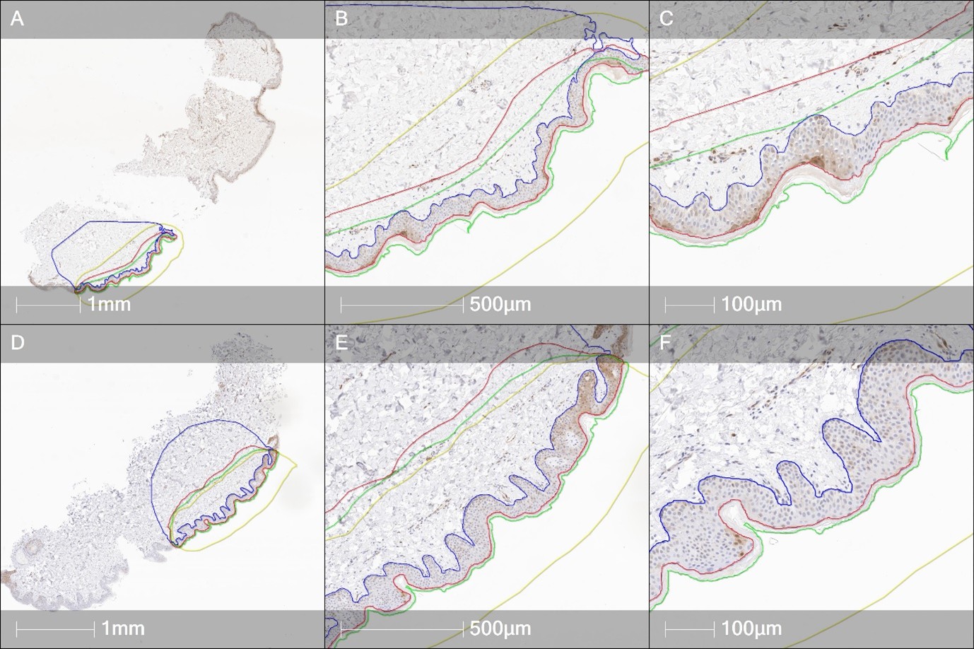

Figure 1: Whole tissue sections (A, D), x10 magnified images (B, E) and x20 magnified images (C, F), all with nested training region annotations for the tissue classes. Classes are shown in ascending order based on the hierarchy used for classifier training.
2 – Capture Staining Variance and Tissue Heterogeneity in Your Training Set
No two tissue samples are identical. Staining intensity can vary between samples, and tissue morphology can present a range of characteristics that a classifier must be able to handle.
It is therefore important to ensure that any staining variance and tissue heterogeneity is well represented in the annotations for the training set. Doing so significantly improves the generalisability of the classifier when it is applied to the broader study sample cohort — meaning it performs reliably across samples it has not previously seen.
3 – Choose the Right Training Resolution
Training the deep learning classifier at a resolution of 1 micron per pixel (x10 magnification) provided a good balance between accuracy and contextual information, enabling precise segmentation of the skin into its Epidermal, Dermal, and Keratinized layers.
In this study, optimal classification was achieved at approximately 7,000 training iterations, which was completed within a few hours. This made for a relatively quick algorithm development cycle — though it is worth noting that training times can be considerably longer for studies with larger tissue sample sets or more complex tissue morphology.
Results: classifier performance
The trained HALO AI DenseNet v2 algorithm was applied to IHC-stained skin images and demonstrated precise layer-by-layer classification across the tissue sections.
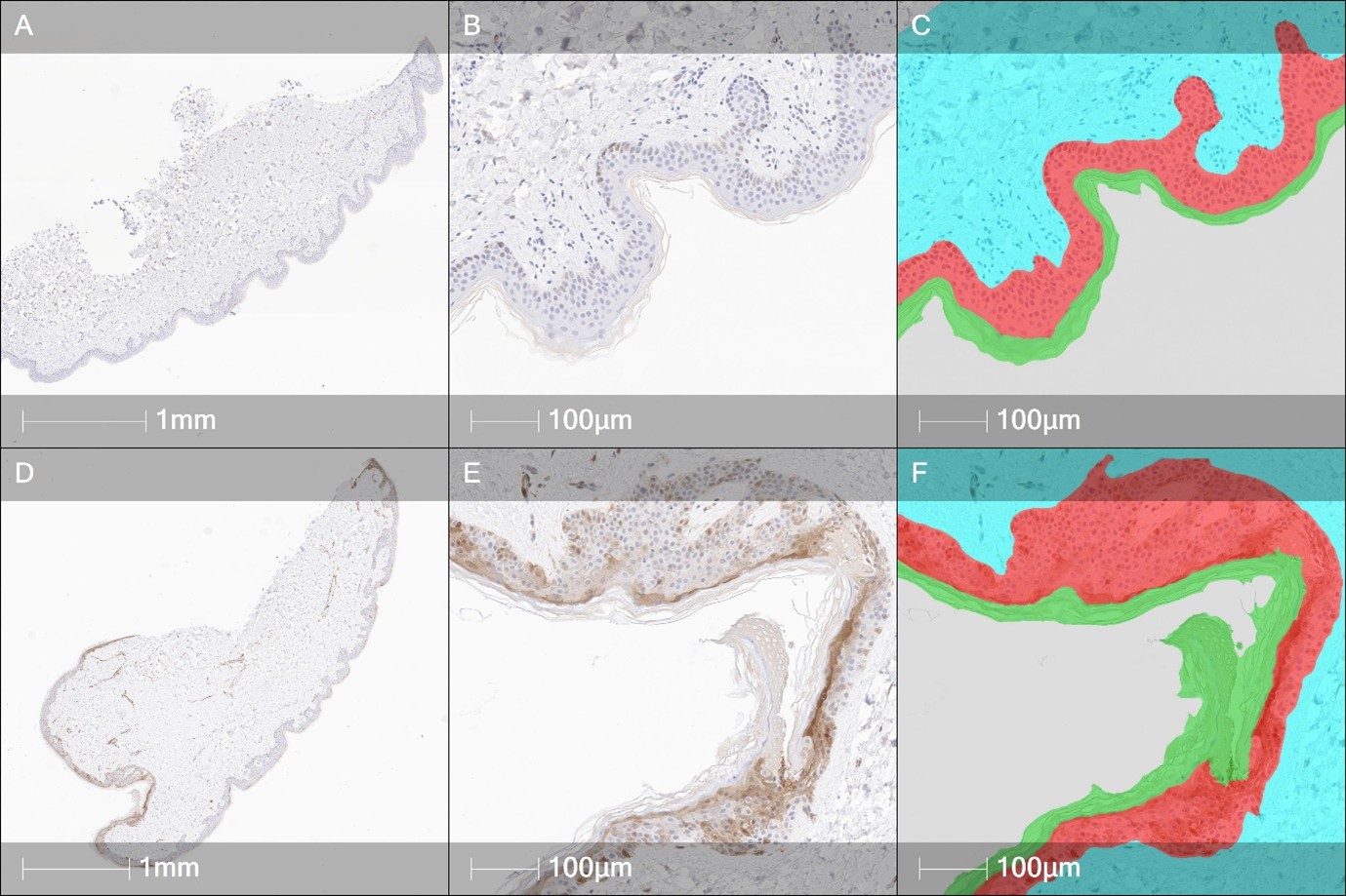

Figure 2: Original whole tissue sections (A, D), x20 magnified original images (B, E), and x20 magnified images with classifier overlay (C, F), showing the classified tissue layers.
Conclusion
The DenseNet v2 classifier algorithm in HALO AI is a powerful tool for skin tissue segmentation. When combined with high-quality tissue sections and carefully constructed training regions, it enables rapid and effective segmentation and analysis of IHC-stained samples.
Getting the foundational steps right — particularly annotation quality, representative training data, and appropriate image resolution — is what makes the difference between a classifier that works well in isolation and one that generalises reliably across an entire study. Done well, this approach can meaningfully accelerate the generation of high-quality, spatially resolved biomarker data.
How OracleBio can support you
Why work with OracleBio?
Stay up to date with OracleBio
Sign up to our mailing list to stay informed about news, webinars, events and more